
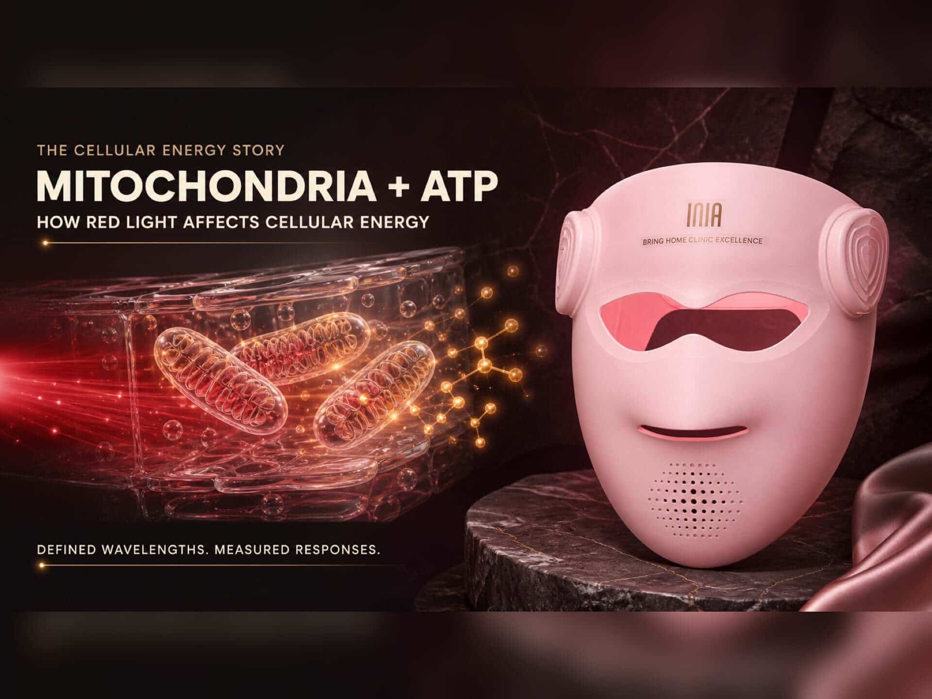
Red and near-infrared light can change how mitochondria manage cellular energy. Studies have measured changes in mitochondrial respiration, membrane potential, ATP, and cell signaling after defined light exposures.
Table of Contents
How Red Light Reaches the Cell’s Energy System
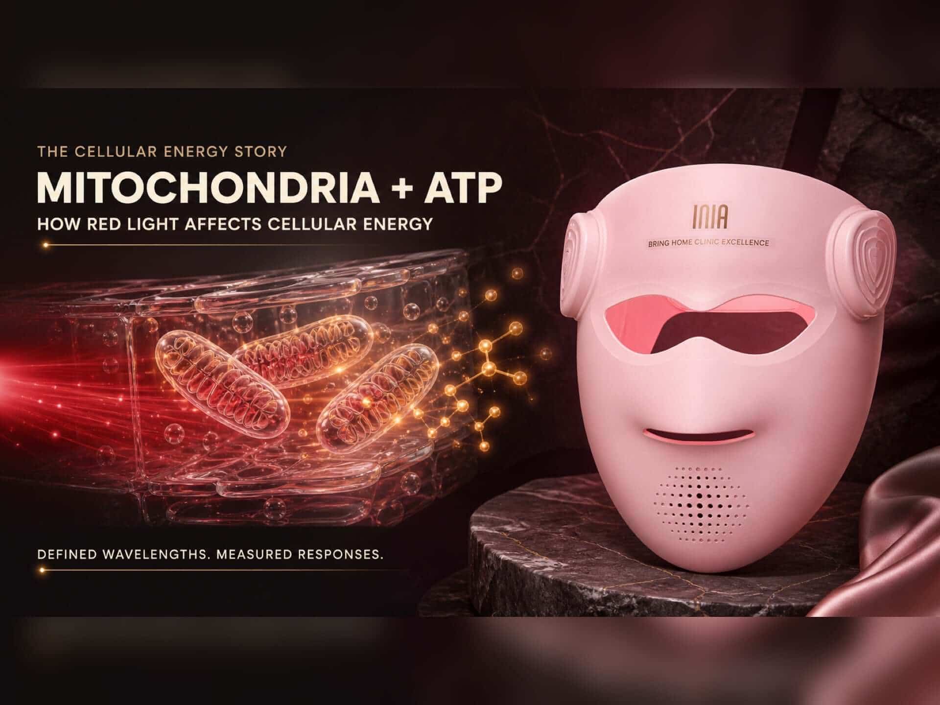
Mitochondria turn nutrients and oxygen into adenosine triphosphate, or ATP. Skin cells use ATP for barrier renewal, protein production, membrane transport, and responses to environmental stress.
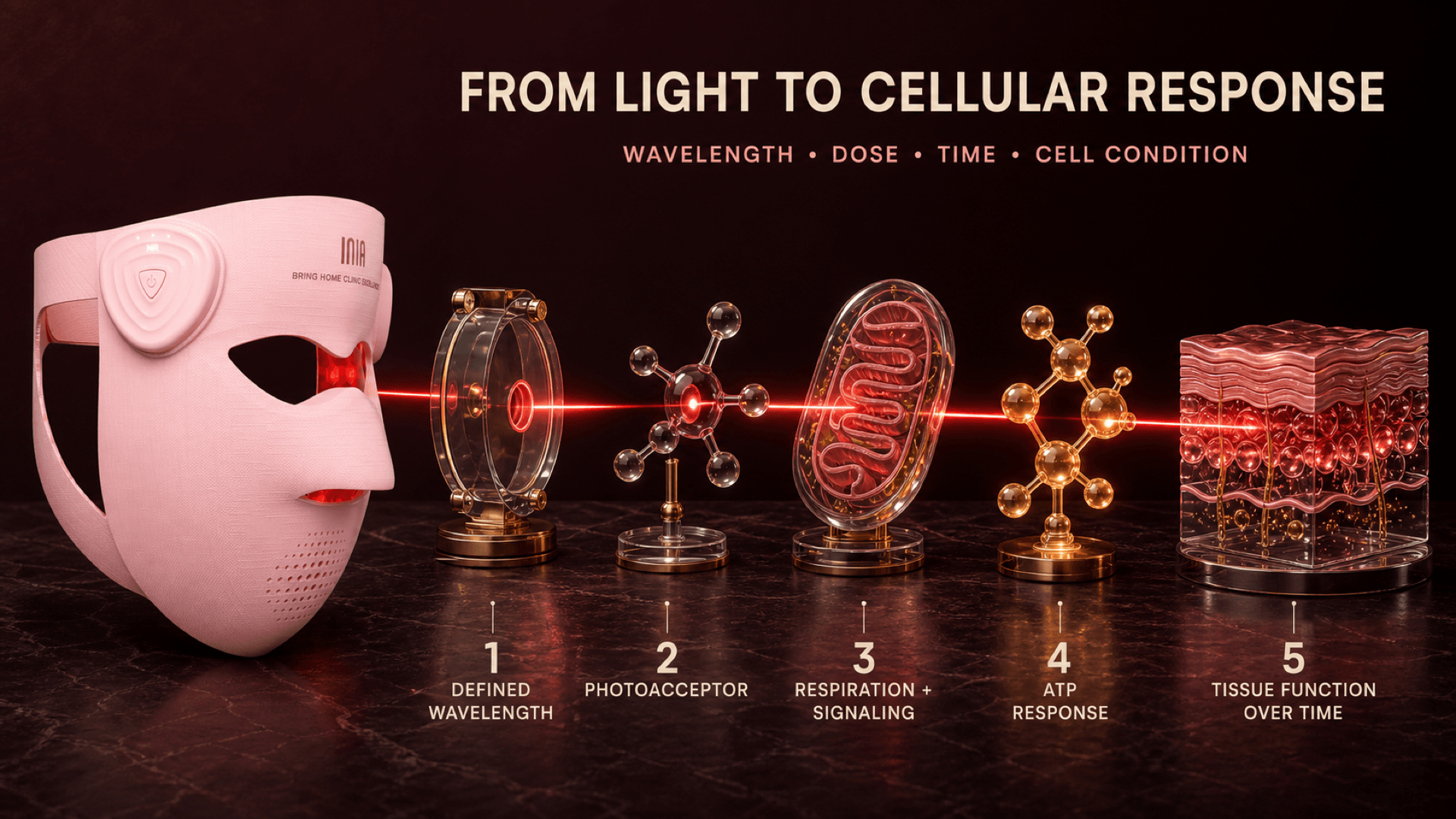
Red and near-infrared photons can reach light-sensitive molecules inside cells. One widely studied pathway involves cytochrome c oxidase, an enzyme in the mitochondrial electron transport chain. Light absorption can change electron transport, membrane potential, nitric-oxide signaling, ATP, and communication with the rest of the cell.1
- A defined wavelength reaches the tissue.
- Cellular photoacceptors absorb the light.
- Mitochondrial respiration and signaling change.
- ATP and related signaling molecules respond.
- Repeated cellular responses can influence tissue function over time.
This cellular response is called photobiomodulation. Wavelength, dose, treatment time, and cell condition shape the response.
What the Research Has Measured
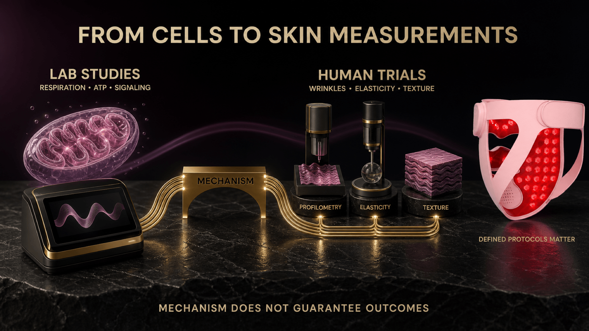
Laboratory studies examine the cellular steps directly. Human trials test visible or measurable changes in skin.
Red light changed mitochondrial activity in human skin cells
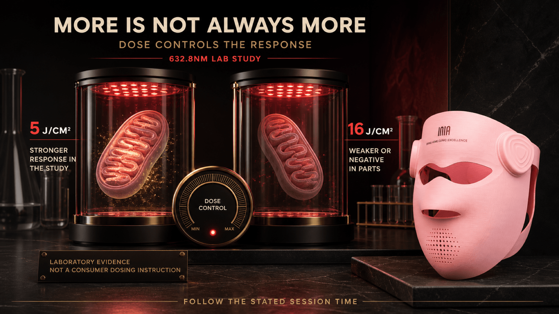
One laboratory study exposed normal and stressed human skin fibroblasts to 632.8nm light. A dose of 5 J/cm² increased mitochondrial membrane potential, ATP, cyclic AMP, and intracellular calcium in several stressed-cell models. A 16 J/cm² dose produced a weaker or negative response in parts of the experiment.2
The practical lesson is clear: dose controls the response. Longer exposure and higher energy do not automatically create a larger cellular benefit.
Red light affected energy metabolism in epidermal cells
A study published online in 2025 compared wavelengths in human keratinocytes. Red light increased oxygen consumption and activated an AMPK-dependent pathway involved in mitochondrial fatty-acid oxidation. The effect remained measurable for up to 48 hours under the study conditions.3
This connects red light with a specific metabolic response in skin cells. It also shows why wavelength matters: different parts of the visible spectrum produced different cellular effects.
Human trials connect defined LED protocols with skin measurements
A randomized, placebo-controlled split-face study tested 633nm red light, 830nm near-infrared light, their combination, and a sham treatment. After four weeks, researchers reported improvements in wrinkle and elasticity measurements in the active groups and documented changes in collagen and elastic fibers.4
A home-use split-face study using 637nm and 854nm LEDs also reported higher elasticity on the treated side after six and eight weeks.5
What Mitochondrial Research Means for Your Skin Routine
“Mitochondrial charging” is a simple name for a measured biological response. It does not tell you whether every red-looking device delivers the same wavelength or dose. Use four practical checks when comparing a mask:
| What to check | Why it matters |
|---|---|
| Exact wavelengths | Biological responses depend on wavelength. |
| Stated session time | The delivered dose changes with exposure time. |
| Consistent facial coverage | Uncovered areas receive a different light exposure. |
| A routine you can repeat | Human trials evaluate a series of defined sessions over time. |
A red-looking device does not reveal its wavelength, dose, or near-infrared output. Check the specifications and follow its current instructions. Adding extra minutes changes the dose and moves away from the stated protocol.
How GLOW 4D Turns the Science Into a Defined Routine
The INIA GLOW 4D Wireless LED Light Therapy Mask combines 630nm red light with 850nm and 940nm dual near-infrared light in its Anti-aging Mode. The stated session time is 10–20 minutes.
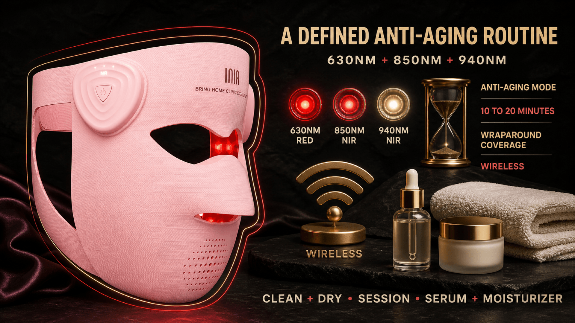
The 4D silicone shape wraps across the face, jawline, lower face, and under-chin area. Wireless operation supports movement during the session. These features support consistent coverage and repeatable use.
Begin with clean, dry skin, complete the stated session, then continue with serum and moisturizer unless the current device instructions say otherwise.
Defined Wavelengths and Timing Make the Science Usable
Red and near-infrared light can affect mitochondrial respiration, ATP, and cellular signaling when wavelength and dose are defined. Choose a mask with transparent specifications, clear timing, stable coverage, and a routine you can maintain.
Frequently Asked Questions
Does red light therapy increase ATP?
Specific red-light exposures have increased ATP or related mitochondrial measurements in laboratory studies. Wavelength, dose, cell condition, and timing determine the response.
Can you feel your mitochondria being charged?
Mitochondrial signaling occurs below the level of sensation. Warmth or brightness describes the wearing experience and does not measure ATP production.
Does a longer red light session create more cellular energy?
Not necessarily. Dose-response studies show that more light can produce a different response. Follow the stated session time.
Why combine red light with near-infrared light?
Both wavelength ranges appear in photobiomodulation research and human skin protocols. A defined combination delivers the visible red and near-infrared portions of the selected program in one session.
Does invisible near-infrared light still work when I cannot see it?
Near-infrared light sits outside normal human vision. Visibility tells you what your eyes can detect; the device specification tells you which wavelengths it emits.
References
- Hamblin MR. Mechanisms and Mitochondrial Redox Signaling in Photobiomodulation. Photochemistry and Photobiology. 2018.
- Zungu IL, Hawkins Evans D, Abrahamse H. Mitochondrial responses of normal and injured human skin fibroblasts following low level laser irradiation—an in vitro study. Photochemistry and Photobiology. 2009;85(4):987–996.
- Herrera MA, et al. Mitochondrial fatty acid oxidation is stimulated by red light irradiation. FEBS Letters. 2026;600(1):20–38.
- Lee SY, et al. A randomized, placebo-controlled split-face clinical study on LED phototherapy for skin rejuvenation. 2007.
- Ng JNC, Wanitphakdeedecha R, Yan C. Efficacy of a home-use light-emitting diode device at 637 and 854-nm for facial rejuvenation: A split-face pilot study. Journal of Cosmetic Dermatology. 2020;19(9):2288–2294.